Blood Flow Through the Heart Page
View this YouTube video to supplement textbook reading.
Preview original video for next week's lesson on EKG.
Diagram of the heart that should be memorized for national boards.
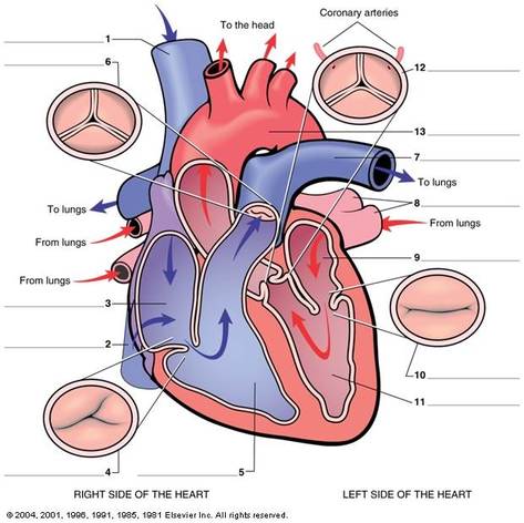 Diagram of the Heart
• Arteries - carry oxygen-rich blood away from the heart to the body’s tissues.
• Veins - take oxygen-poor blood back to the heart.
• Capillaries - are small thin blood vessels that connect the arteries and the veins. This is where the oxygen/carbon dioxide exchange takes place.
1.Deoxygenated blood
enters the heart through two large veins, the inferior and superior vena cava #1
and # 2, emptying into the right atrium. #3
2. Blood flows from the right atrium #3 into the right ventricle #5 through the
tricuspid valve. #4
3. Blood then leaves the right ventricle #5 and the heart through the pulmonic
valve #6 into the pulmonary artery #7 (only artery to have oxygen poor blood) and to the lungs.
4. Oxygenated blood returns from the lungs to the heart via the pulmonary veins #8 (only veins to have oxygen rich blood) into the left atrium #9.
5. From the left atrium, blood flows to the left ventricle #11 through the mitral valve #10.
6. From the left ventricle blood leaves the heart through the aortic valve #12 into the aorta #13 and to the body.
Exercise One - Identify numbers 1-13
Briefly describe oxygenated and deoxygenated blood flow through the heart.
Briefly describe oxygenated and deoxygenated blood flow through the heart.

Diagram of the Heart
Task Three - Diagram Assignment
The student will create an original diagram of the heart and trace the pathway of the blood through the heart with appropriate labeling of 13 major points of anatomy; the oxygenated and deoxygenated blood flow through the heart will be clearly distinguished. The diagram may be an actual representation of the heart, a smartchart, or diagram of your choosing. See example below.
Submit this assignment for the class to view via dropbox or in the assignment space provided.
After submission, assignments will be posted to the website for review by members of the class.
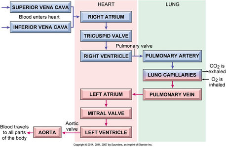 Diagram of Blood Flow through the Heart
Sample Diagram of Blood Flow through the Heart